Role of radiologist reading mammograms ever-changing
A radiologist’s role in reading mammograms for breast cancer diagnosis has come a long way since the days of confining themselves to interpreting mammograms and dictating reports to the referring physician.
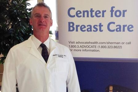
Now on the frontlines, a radiologist remains with the patient almost every step of the way from screening through diagnosis, says Dr. Stephen Grossman, radiology and breast specialist at the Center for Breast Care at Elgin-based Advocate Sherman Hospital.
Having spent the last 11 years at Sherman Hospital, Grossman and the breast care team play an integral role in interpreting breast imaging studies, delivering results and creating a plan for patients who receive abnormal mammogram results.
Soon, when a woman comes in for her screening appointment, which is read the same day by a radiologist board-certified in mammography, she can receive a telephone call with her results the very next day.
If the results come back normal, patient-anxiety is short-lived, Grossman says, but when an area of concern is identified or results are suspicious for breast cancer, patient stress-levels rise, and the goal is to promptly begin the process of obtaining a diagnosis and keep the patient as informed as possible.
Breast cancer is a tricky thing, says Grossman, especially when some findings that look benign turn out to be abnormal, while other results look suspicious, and end up being nothing at all.
Grossman sat down with Sherman Hospital Public Affairs and Marketing Coordinator Lawerence Synett to discuss the radiologist’s role in mammograms and delivering the news.
Synett: What happens after a woman receives a mammogram?
Grossman: There are three possible results from a screening mammogram: negative, if it is completely normal; benign, if there are some findings but they are clearly benign; and incomplete, if we need to further evaluate. Comparison with previous mammograms is very important to insure that no subtle changes are present.
If we need to further evaluate, it becomes a diagnostic study, which usually involves additional mammogram views of the area of concern, and frequently an ultrasound. Some things show up more clearly during an ultrasound, and breast ultrasound is often required to evaluate whether a biopsy is needed. Even if the findings don’t appear suspicious, we may need to keep a closer eye on things to make certain that no changes are happening. In this case we may recommend a repeat study in six months rather than a full year. Women are informed of their results and are provided a written summary before they leave our facility.
At the Center for Breast Care, women are involved in any and all decisions regarding their breast health. Women often ask what I would recommend if they were a member of my family. I respond with honesty and compassion. If am not completely convinced that everything is OK, I will recommend a needle biopsy. If I am 98-percent sure it is OK, just to be on the safe side, I recommend a six-month follow-up. I also make it clear that if waiting causes too much stress, I am happy to offer a needle biopsy to make absolutely certain that there is nothing wrong.
Synett: How do you proceed if a biopsy is needed?
Grossman: If a biopsy is needed, we sit down with the woman and explain everything. If it is something best seen on the mammogram, we generally do a stereotactic needle biopsy. This procedure requires only local anesthetic. On a special mammogram table, the woman lies on her stomach, and we can place the biopsy needle to the exact spot. The actual procedure lasts just a few minutes. If it is something we can see using breast ultrasound, we can perform a similar needle biopsy even more comfortably.
Breast MRI is the most sensitive test that we have for finding the smallest and hard to find breast cancers. More recently, we have started using breast MRI to guide needle biopsy, to sample suspicious findings found only with MRI. In this way, we recently diagnosed a 2 mm breast cancer, which is the smallest we have yet to find.
All of our needle biopsies are done without any stitches, through a 3-mm nick in the skin. There are very few complications. Because needle biopsy has become so accurate, the need for a six-month follow-up is now less common.
For the majority of women, needle biopsy confirms that no cancer is present. For those women where we find something wrong, needle biopsy allows a surgeon the opportunity to accurately counsel and plan for a single definitive procedure.
Synett: If the results are serious, how are the results communicated to the patient and how is a plan put in place?
Grossman: After a patient has a biopsy, we discuss how she would like to receive the results from us. Usually we have the pathology results in 48 hours. We coordinate in advance with the referring physician to arrange for prompt surgical referrals should it become necessary. We understand how stressful it is to wait, so we have developed as much efficiency as possible.
Even when the news is bad, there is tremendous hope because treatment is now so effective. When caught at an early stage, breast cancer is very treatable with a high success rate. All of us at the Center for Breast Care are proud to participate in this vital part of a woman’s health care needs.
Related Posts


Comments
About the Author

health enews staff is a group of experienced writers from our Advocate Health Care and Aurora Health Care sites, which also includes freelance or intern writers.